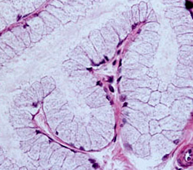
TUBULAR MUCOUS GLAND – 3
Detail of a sublingual salivary gland, which is formed by numerous mucous tubules.
Most of the upper figure is occupied by a twisted and branched mucous tubule highlighted in light blue and the tubular lumen in pink. It is surrounded by portions of neighboring tubuli highlighted in a darker tone of blue and by connective tissue in red. Notice at the lower right corner an arteriole (light green) and a venule (darker green).
Notice the secretory product within the wide lumen of the large tubule.
No conducting part of the gland (excretory ducts) are present in the figure.
Secretory cells
In the lower image observe a slighly enlarged image of the same tubulus. The cytoplasm ot its columnar or pyramidal secretory cells does not stain orange with eosin but in a slightly bluish color, due to the presence of many acidic groups in the molecules of the secretory product kept in small vesicles. The cytoplasm appears somewhat vacuolated and heterogeneous (i.e., not very “smooth”).
Three secretory cells become highlighted in blue-violet after moving the mouse or clicking. The nuclei of the secretory cells are elongated and appear to be “squeezed” against the base of the cell. They stain strongly with hematoxylin because contain mainly condensed chromatin. Some nuclei turn red when using the mouse or clicking.

Sublingual salivary gland. Staining: hematoxylin and eosin. Small magnification.